A dentigerous or follicular cyst is defined as a cyst that originates from the separation of two specific components of a developing tooth. A cyst is basically an enclosed sac within tissue containing either fluid, air, or semi-solid materials. In this case, the follicle separates from the developing crown of a tooth, and usually a collection of fluid develops in the cystic space.
Dentigerous Cyst Case #1

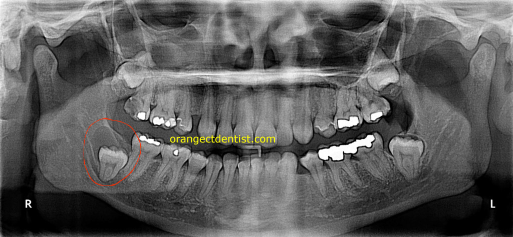
Dentigerous Cyst on the lower right with an impacted wisdom tooth. This female patient was from West Haven, CT. X-ray and referral for surgical treatment by Dr. Nicholas Calcaterra.
The panoramic x-ray above is from a 28 year old female. She had never had a panoramic x-ray before. The red circle shows an unerupted lower right wisdom tooth with a cyst around it. The tooth along with the cyst was removed surgically and the patient recovered quite well with no significant long term complications.
Description and Treatment of Follicular Cysts
This type of cyst is the most common type of developmental odontogenic (tooth associated) cyst, making up 20% of all of these types of cysts of the jaw. They most often occur with impacted third molars (wisdom teeth) as seen in the radiograph above. They are occasionally associated with supernumerary teeth or odontomas. Most of these cysts are seen in patients between the ages of 10 and 30. Small ones are usually without symptoms and only discovered upon routine digital radiographs. If undetected, they can continue to grow in size, leading to a painless expansion of the area and even visible facial asymmetry.
The most common treatment for these cysts is enucleation (removal of the object without cutting directly into it) along with removal of the enerupted tooth/teeth. This is typically performed by an Oral Surgeon in an outpatient setting under anesthesia. Larger ones may require a different technique called marsupialization. The prognosis for these is excellent and recurrence is infrequent.
References
All clinical examples are from the dental practice of Dr. Nicholas and Calcaterra. Other data adapted from: Neville, Brad W.. Oral & Maxillofacial Pathology, 2nd Edition. Saunders Book Company
